
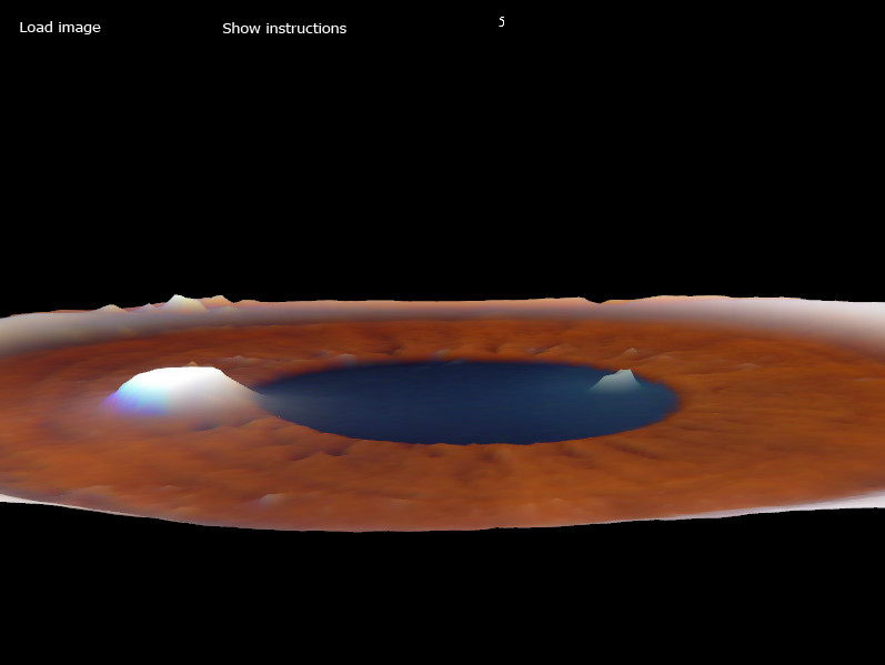
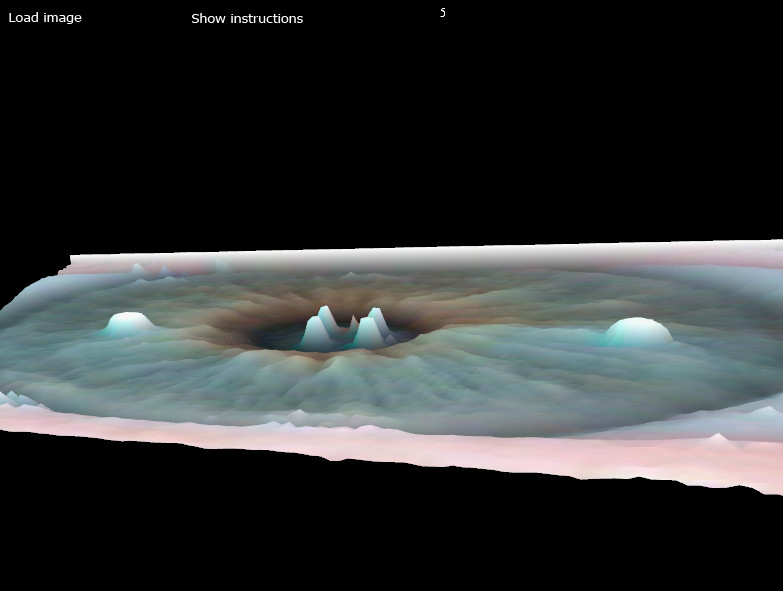
This program was designed to assist iridological researchers and practitioners to review the human eye using 3D landscape view. The most important aspect of viewing the eye in 3D landscape view is in determining the iris relief which can be very difficult analyze using standard photo image. Please note that the 3D iris View project is for research and development only.
The Relief of the Iris Surface
The autonomous nerve wreath plays an important role in iris topograpy, for the wreath is usually elevated over the other areas of the iris. It is advantageous to examine the relief of the iris with side illumination, which makes all the structures more distinctive.
All aforementioned components and features found throughout the iris provide pertinent information for reaching iridological conclusions. Irises of every person are very unique. In fact, the iris is considered to be an even more reliable distinctive “marker” of an individual than the fingerprint.
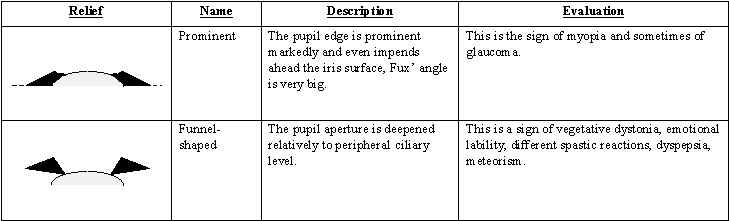
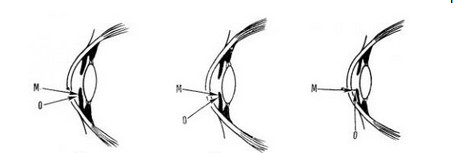
An iris relief is the profile of the iris surface, which is never flat or plain but a continuity of prominences and depressions. The iris surface is raised from the pupil center to the prominence of the autonomous wreath, and the shape of the wreath varies greatly. From this level the surface of the ciliary belt descends as a slight slope to the external edge of the iris. The best method of examining the iris relief is the slipping beam method which uses side illumination. Evaluating the iris relief gives us an opportunity to examine reserve and self-protecting capacities of the organism.


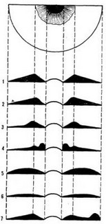
R. Bourdiol further described two other relief forms:
I – normal
2 – disc-shaped
3 – flattened lateral
4 – crater form
5 – rounded, thickened
6 – flat
7 – locally deformed

Velchover data
Interesting information in terms of morphogenesis are differences relief iris. The study gives us relief data protection and backup capabilities of man. It is best to study the relief of side light iris, by moving the beam. The surface of the iris does not look smooth and flat, but a conglomeration of bumps and depressions, something resembling volcanic craters. From the center (or pupil) iris surface rises to an elevation of independent rings, the shape of which is very variable. G edge surface elevation ciliary zone descends in the form of a gentle slope to the outer edge of the iris. There are several types of terrain. We describe the seven varieties of terrain and interpretation G. Jausas (1974):
Types of relief iris. I – normal, 2 – disc-shaped, 3 – flattened lateral 4 – crater form 5 – rounded, thickened, 6 – flat 7 – locally deformed.
1 – Normal. Have an average size of the ring tops autonomous and uniform internal slopes. Indicates the balance of vitality and good prognosis in diseases.
2 – Disc-shaped, etched characterized pupillary zone in the middle. Occurs in hypertension, bradycardia, sweating and diarrhea.
3 – Flattened lateral, etched characterized slope ciliary zone, demonstrating the sympathetic hypofunction.
4 – Crater-form. Different steep slope the foremost pupil zone. Found in endocrine and humoral disorders.
5 – Rounded, thickened. The surface of the iris as a swollen, Fuchs angle (formed by the pupillary zone and self-ring) is missing. Occurs in hypertension and polyphagia.
6 – Flat. Characterized by the complete disappearance of the independent rings. Indicates a low level of resistance and poor prognosis in the case of serious illness.
7 – Locally deformed.. Indicates the presence of severe chronic illness.
Each ophthalmologist is well known that the age of iris tissue is undergoing significant changes. Often it concerns the rear leaf pigment. Is destruction pigment border of the pupil. Changes in mesodermal tissue is reduced to its thinning, compaction and loss of transparency, resulting in changing the color of the iris and its relief. Color begins to fade, mostly gray-blue, relief – smooth.
Radii Solaris Differentiation
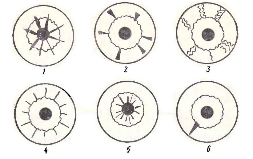
Types of toxic radiance by K. Voig.
1: Asthenic Radial Crack 2: Hyperemic Sulci 3: Stepped Radial Sulci 4: Peripheral Sulci 5: Sulci of the pupillary area 6: Cracks of the autonomic nerve wreath
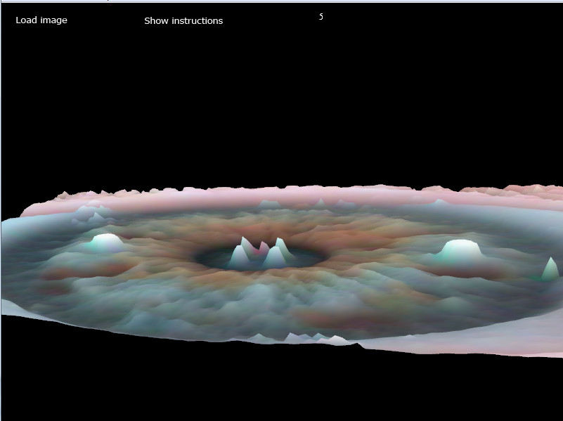
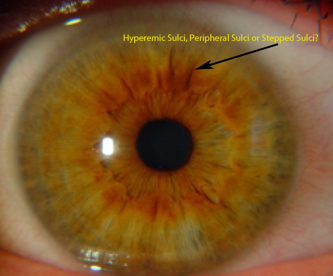
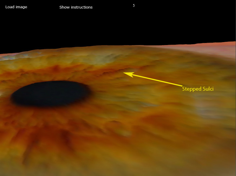
The Stepped Radial Sulci can be difficult to differentiate from the Asthenic Radial Crack, Hyperemic Sulci and Peripheral Sulci. The Stepped Radial Sulci is often seen in the ciliary belt, can be intermittent or echeloned ( type of diffraction grating used in spectroscopy consisting of a series of plates of equal thickness arranged stepwise with a constant offset ). Using 5X – 10X 3D surface view makes it possible to differentiate Stepped Radial Sulci:
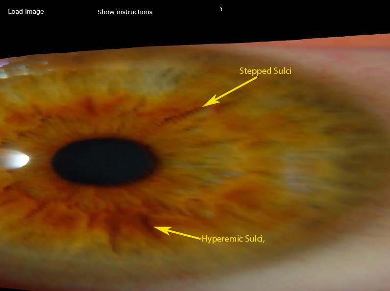
5X – 3D Surface View
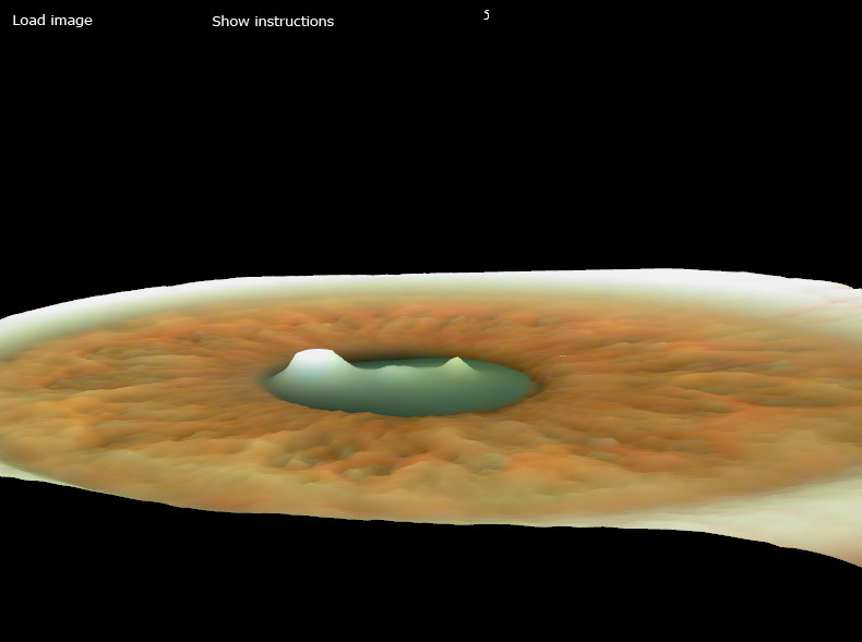
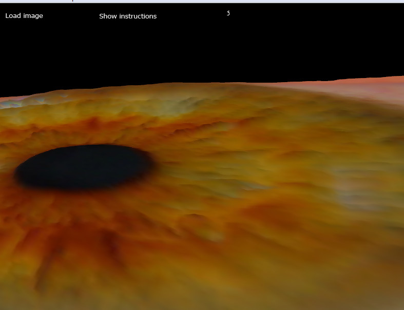
5X – 3D Surface View Zoomed
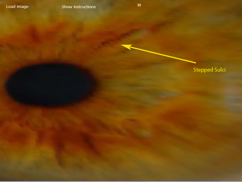
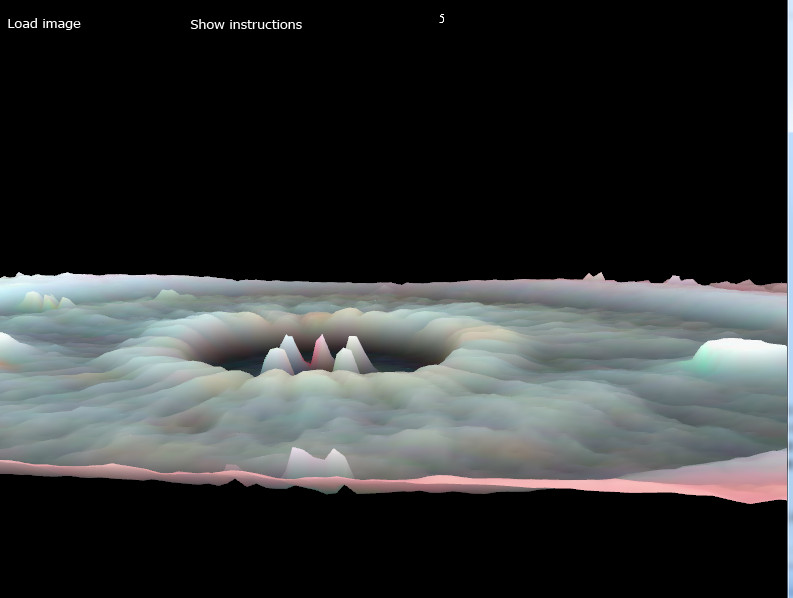
10X – 3D Surface View Zoomed
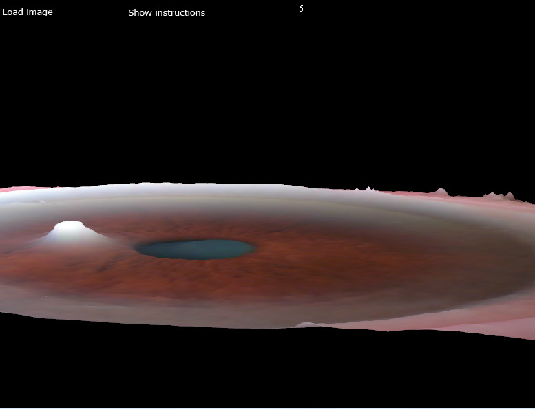
10X – 3D Surface View Zoomed Angle View
The stepped radial sulci is typical for sclerotic changes in tissues. The arrangement of sulcus on the bottom of a lacunae is indicative of irreversible changes in the organ (such as the kidney area in nephrosclerosis).