Clinical iris & pupil analysis
Photograph both irides. Read them back as measurements.
The Iridology Analysis Toolkit measures the pupil margin, the collarette and the iris zones from your own captures, and writes the numbers into a report you can hand to a patient.
Everything runs on the machine in front of you. No cloud, no upload, no subscription — a licence key is checked once, and after that the toolkit works offline indefinitely.
Patient names, ages, images and scan records are stored on the device only. Licence activation sends a machine ID and a key — never a patient identifier.
- Reference circle a circle of the pupil margin's own median radius
- Collarette (ANW) the autonomic nerve wreath, measured as a ratio to the iris
- FLAT margin curving inward — here 7.4%, the Moderate band, in the lower-nasal zone
- PROT margin bulging outward — here 4.2%, the Mild band, in the upper-nasal zone
- Version
- 1.5.0
- Platforms
- Windows 10+
Android 8+, iOS 14+, macOS - Languages
- 26 languages
29 locales - Free trial
- 14 days or
20 analyses
What it measures
The right eye is captured first (OD), then the left (OS) — the standard clinical iridology order. Every figure below is a geometric measurement taken from your own image, with a stated threshold behind it.
- P/I ratio
- The pupil diameter as a percentage of the iris diameter. Both are measured in the same image, so the ratio does not shift with camera distance or magnification. 20–30% is the normal band for an adult indoors, and the reading is also compared against an age-specific reference range.
- Zone findings
- Flattenings (FLAT) and protrusions (PROT) of the pupil margin, each assigned to one of eight iris zones and graded Within limits, Mild, Moderate or Significant by its percentage deviation.
- ANW ratio & asymmetry
- The collarette ring measured against the iris diameter: under 25% reads Spastic, 25–35% Normal, over 35% Atonic. The OD/OS difference is reported too, and one eye Spastic against the other Atonic is flagged as a Functional Frustration pattern.
- Decentration
- How far the pupil centre sits from the iris centre. Past 5% the direction is named as well — nasal, temporal, frontal, basal or a diagonal between them — and the angle is reported in degrees.
- Ellipseness & form
- The pupil's minor axis as a percentage of its major axis, so a true circle scores 100%. Below 95% the ellipse orientation is classified into a form type: horizontal, vertical or diagonal oval, chord-like, or irregular.
- Anisocoria
- The difference between the two P/I ratios. Under 2% is within normal limits; 4% or more raises a TBI research-observation flag in the results and the PDF.
4 severity bands
Two engines, and only one of them can overrule
Every image goes through a classical pixel-based pipeline — circle detection, radial sampling, boundary-point analysis at full resolution — and through an ONNX neural network on a 224×224 crop centred on the iris.
The model contributes one validated figure: the P/I ratio, at R² = 0.856 and MAE 1.17% over 712 Bexel eyes. Zone findings, ANW, decentration and ellipseness come from the classical pipeline alone. The ML PI Agreement line in the report means agreement on the ratio — it does not validate the other measurements, and the toolkit says so rather than implying otherwise.
Geometry suggests, the practitioner confirms
The Velchover ANW wizard pre-ticks patterns whose geometric signature matches the detected sector deviations. Those pre-ticks are suggestions. Only patterns you review and confirm reach the record, the results screen and the PDF.
Skipping the wizard records nothing at all. The same principle runs through the toolkit: a measurement is never promoted into a clinical finding without a human saying so.
What it looks like in use
Captured on Windows, where the toolkit has its fullest feature set. The features page walks through each of these in detail.
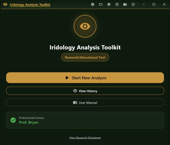
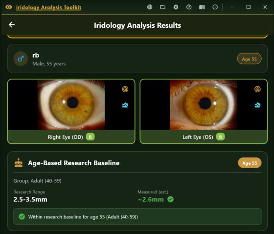
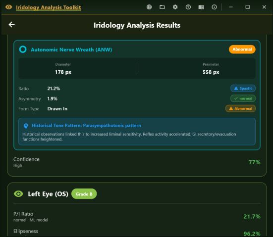
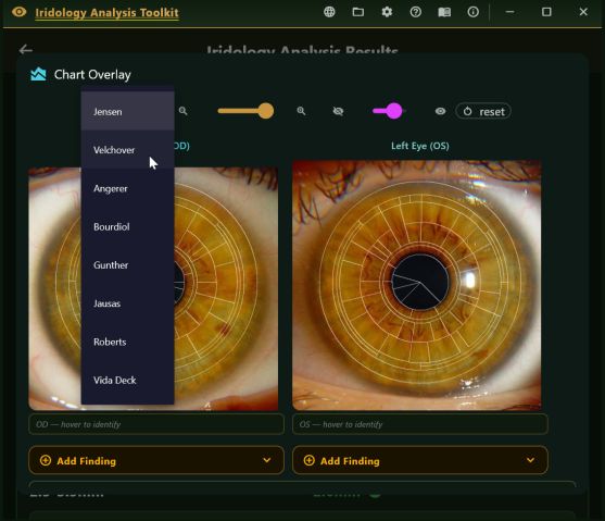
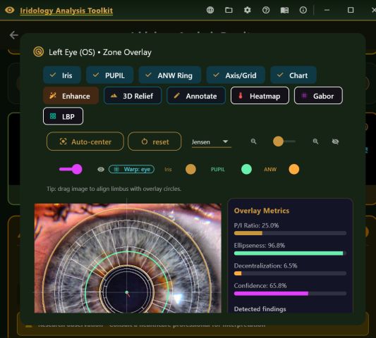
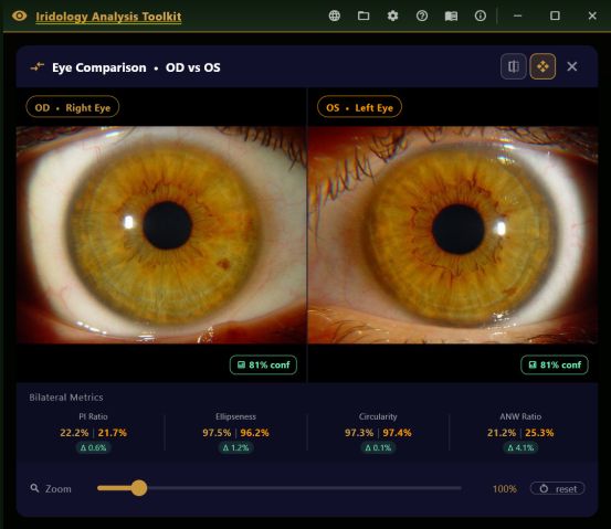
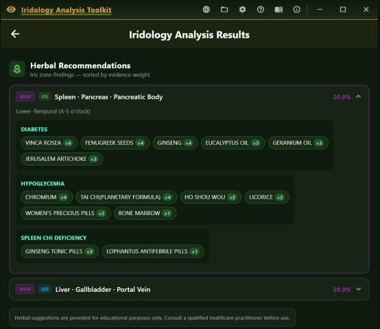
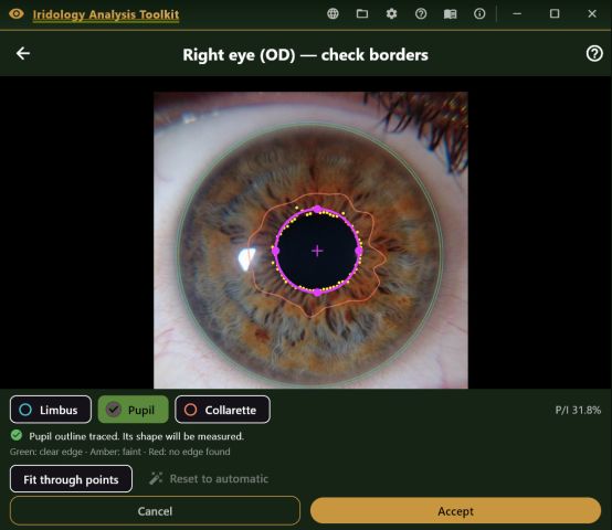
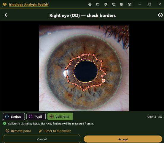
The whole consultation, in one pass
The toolkit follows the same path every time it opens, so the order is never in question and nothing is half-saved.
Patient information
Name, age and sex, plus main complaints and your clinic name. Age drives the age-normalised comparison, and the clinic name is remembered between sessions so you type it once per installation.
Capture, right eye then left
Pick the source per eye: a quality-gated phone camera, a manual shutter, a USB iriscope such as the Dino-Lite, PLR video, or an import from the gallery. The gated modes check sharpness, brightness, contrast, pupil confidence and centring on every frame before letting one through.
Optional constitutional type
With both irides on screen side by side — the moment Deck's system calls for — pick from 34 constitutional types in six groups, or leave it unassessed.
Analyse, then read
The hybrid pipeline runs and the results screen assembles: per-eye metrics, zone findings, the ANW card, decentration and form, the bilateral comparison, and any therapy panels you have enabled.
Annotate and export
Add observer notes, mark the image, overlay a chart, then export a PDF or a plain-text report. The scan saves itself to local history the moment the results finish loading — there is no save button to forget.
Also in the toolkit
26 languages
Interface, clinical wording, the built-in manual and the generated PDF, in 29 locales including Brazilian Portuguese and Traditional Chinese. The chart organ names are translated too.
Scan history and trends
Every analysis is kept in a local database with its full result. From any record, a serial timeline plots P/I ratio, ellipseness and confidence for both eyes across all of that patient's visits.
A research toolkit
Ten specialised tools in their own workspace — crypt and lacuna detection, contraction furrows, heterochromia sectors, Frangi vesselness, GLCM, an iris signature — marked research-only throughout.
The manual travels with it
Five tabs, offline, in every supported language: getting started, capture, reading the analysis, tools and exports, and the therapy and safety sections in full.
Getting it
The free trial runs for 14 days or 20 analyses, whichever ends first, with every feature unlocked and no card required. When it ends you can still open past scans, export reports and browse history — only new analyses stop.
| Tier | Duration | Machines | Notes |
|---|---|---|---|
| Trial | 14 days / 20 analyses | 1 | Every feature unlocked. Reinstalling does not restart it. |
| Standard | 1 year | 1 | Single-machine key for an individual practitioner. |
| Professional | Lifetime | 1 | Single machine, no renewal. |
| Enterprise | Lifetime | Multiple | For clinics and teaching institutions. |
Keys are bound to a machine ID at activation, which needs an internet connection once. After that the toolkit runs offline indefinitely. Android and iOS are licensed through their own stores.
What you need to run it
- Windows (primary) — Windows 10 64-bit, 4 GB RAM, a USB 2.0 port if you are using an iriscope
- Android 8.0 or newer, with USB OTG if you are attaching an iriscope
- iOS 14 or newer
- macOS — installed directly from this site
Cameras it will capture from
A Dino-Lite AM4115ZT or a compatible UVC iriscope over USB, a phone or tablet camera through the quality gate, a manual shutter for macro-lens work, PLR video, or an existing photograph imported from the gallery.
Imported images are copied into a folder the toolkit manages, so your original file is never modified.
What this is, and what it is not
A research and educational tool
The Iridology Analysis Toolkit is for licensed practitioners trained in iridology, natural medicine or a related field. It is not a medical device and has not been approved, cleared or certified by the FDA, CE, TGA or any equivalent authority for diagnostic use.
Every figure it produces — P/I ratio, zone findings, ANW assessment, decentration, ellipseness, anisocoria, age-normative comparison, confidence score and all therapy content — is observational and educational. It is not a diagnosis, and not a substitute for a clinical examination. The practitioner is solely responsible for the clinical decisions made from it.
The measurement and the meaning are separate things
Zone names, organ associations, constitutional types and therapy suggestions are drawn from the historical iridology literature and from the CNRI reference charts. The toolkit measures geometry accurately; it does not test whether the historical correspondence holds. An exact measurement of a flattening is not evidence of what that flattening means, and the two should be kept apart when you read a report.
A reflection can imitate a flattening
A specular highlight sitting close to the pupil border reads as a straightened edge. The toolkit raises a warning on the card, in the PDF and on the stored scan when a reflection falls inside the limit, and no measurement is withheld — but the warning identifies images at risk, not every affected image. A real deformation stays put when the light moves; a reflection travels with it.
Your patients' data stays yours
Names, ages, images and scan records are written to local storage on the device and nowhere else. Nothing is transmitted to CNRI, to a cloud service or to any third party. You remain responsible for making your local storage and your exported reports comply with HIPAA, GDPR, the Australian Privacy Act or whichever law governs your practice.
See the whole thing, measurement by measurement
The features page carries every threshold, every tool and every module — it replaces both the old features page and the old in-depth page.