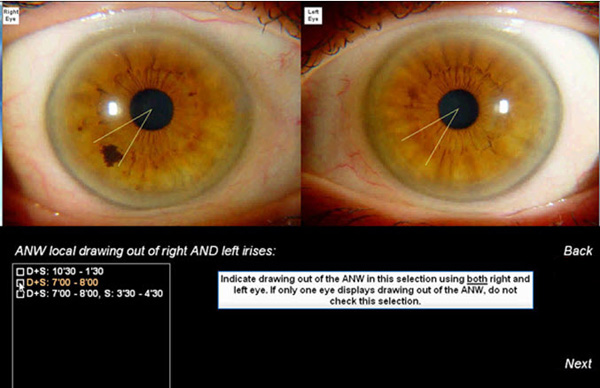
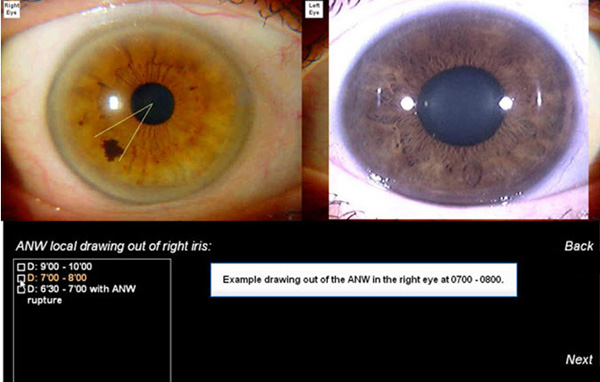
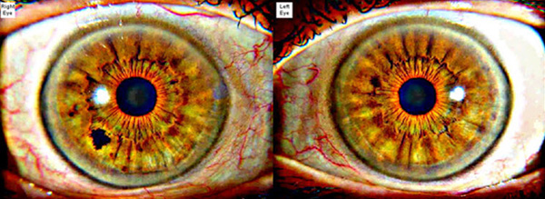
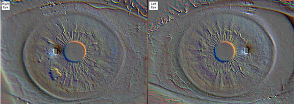
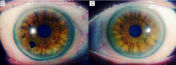
This iridodiagnostic software was developed using several hospital clinical studies accomplished by the famous neurologist Prof. Velchover in collaboration with the University of Peoples’ Friendship in Moscow. Dr Velchover is known as the father of modern scientific iridology. This software was introduced to Medical doctors in 2001. In Russia, Iridology is accepted as official science and taught in several medical universities and institutions.
Key Highlights
1: Iridology Charts
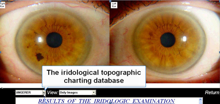
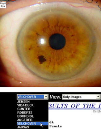
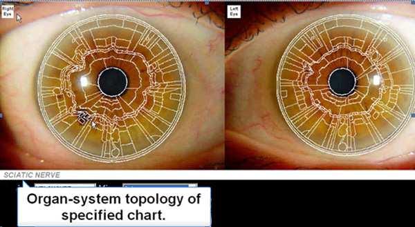
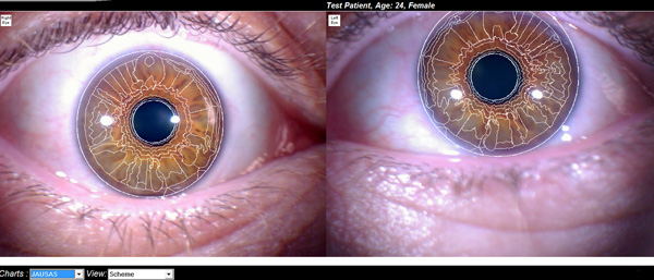
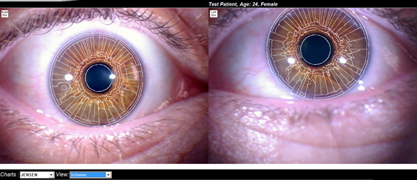
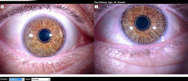
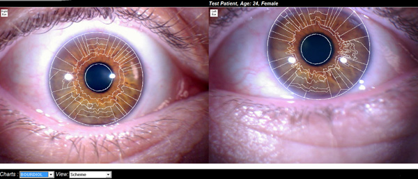
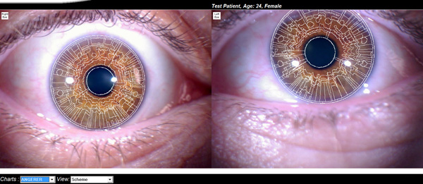
Eight different iridological topographical charts are offered in program. Each specified chart is properly set over each unique eye for clear over-lay use and determining organ and system sectors. Charts available include Jensen, Vida-Deck, Gunter, Roberts, Bourdiol, Angerer, Velchover and Jausas.
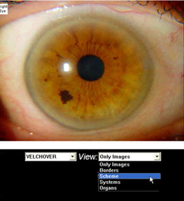
Choose iridological chart and then the View: Scheme box:
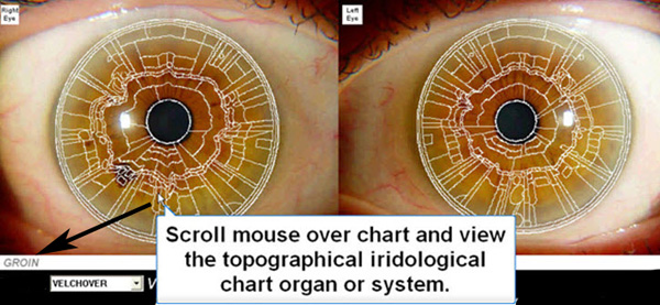
Defining Iris Topography Sections Using Scrolling Mouse-Over Charts
Choice of eight iridology charts precisely applied to the human eye using complex bio-metric algorithm that will automatically detects the pupil, collarette and iris limbus parameters. Currently, there are over 150 unique pupil and collarette signs that can be automatically detected with information applied to final assessment report.
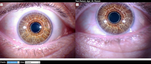
Velchover Iridology Chart
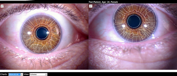
Vida-Deck Iridology Chart
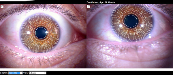
Roberts Iridology Chart
Jausas Iridology Chart
Jensen Iridology Chart
Gunter Iridology Chart
Bourdiol Iridology Chart
Angerer Iridology Chart
2: Automatic Detection of the Eye, Pupils and Collarette
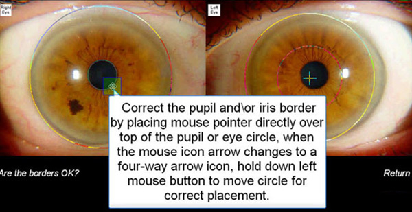
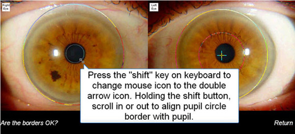
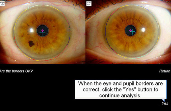
The iridodiagnostic software will attempt to automatically place topographic charts over each eye. If base chart is not correct you can make the required changes in this section manually as shown below:
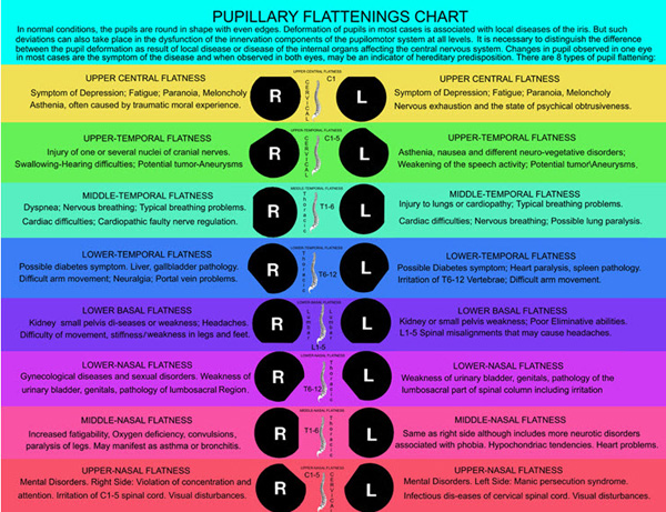
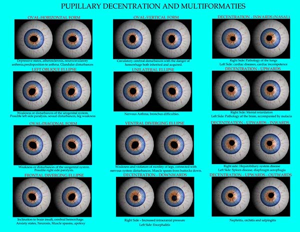
The pupil of the eye and its photo-reaction are visible reflections of the functional state of different brain structures. Information is combined about both afferent (somatic) and efferent (vegetative) components of cranial-cerebral innervation and pupilomotor systems of the eye, and the pupil is an accessible and informative object for evaluating truncus cerebri activity. Pupil reflexes play the primary role in making a diagnosis of many neurological diseases as a part of well-known syndromes: Bernard-Horner, Adie’s, Argylle-Robertson, Parinoud’s etc. Changes in the pupil of color, dimensions, shape, position of center, equality and reflector reactions all have clinical significance. Iridology Analysis Pro can accurately detect pupillary deformations and pupillary decentrations:
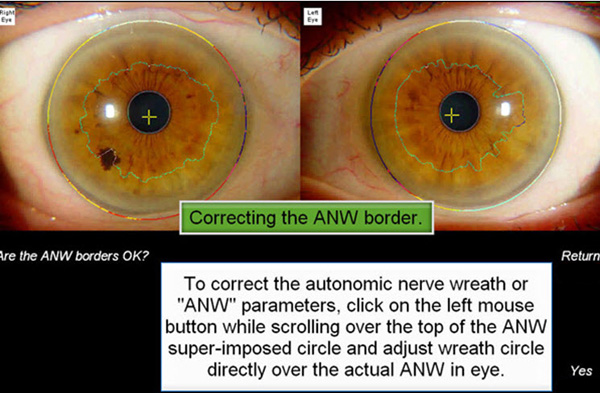
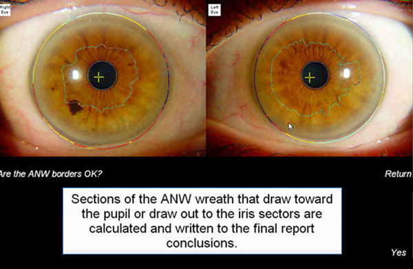
Correcting the Autonomic Nerve Wreath
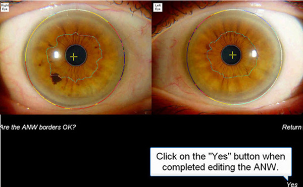
The iridodiagnostic software will attempt via biometrics to automatically place the collarette chart over each eye. If base chart is not correct you can make the required changes in this section manually as shown below:
If required, editing the collarette is very fast to accomplish. Simply click your mouse pointer to undetected sections of the collarette for placement.
3: Choosing Eye Color, Density & Relief
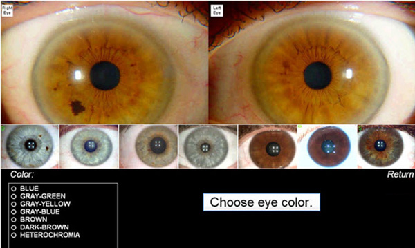
Choosing Eye Color
Samples are shown to assist in choosing correct eye color.
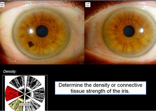
Choosing Density
Simply scroll mouse of the Density Chart to choose the correct density.
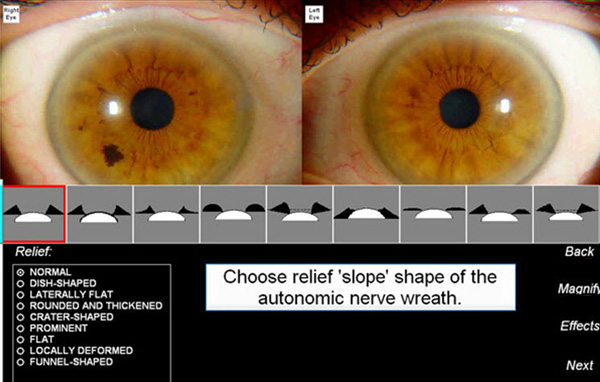
Choosing Relief
Analyzing the iris relief requires a great deal of practice. If the user is unfamiliar with the iris relief shape it is best to check normal and will not be included in final analysis report. We have designed a small program to analyze the eye in 3D to assist in reviewing the iris relief.
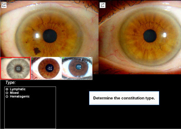
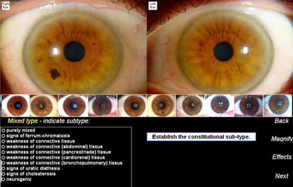
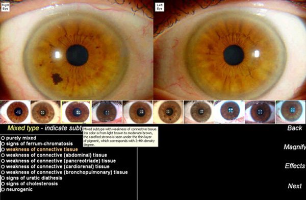
4: Determining Constitution Types
Determining the main constitution and possible sub-types is an important feature in this software.
Choose the correct constitution type and Sub-type.
Scroll over constitutional sub-types sample windows in software to display more detailed information.
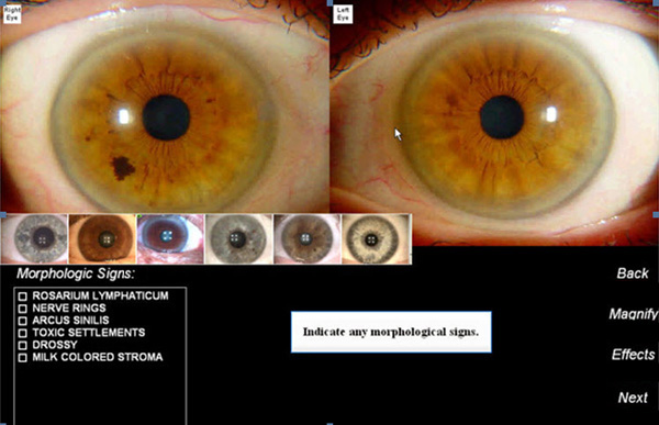
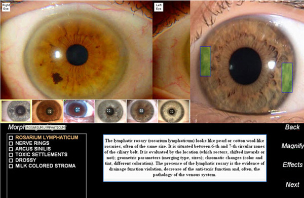
5: Determining Morphologic Signs
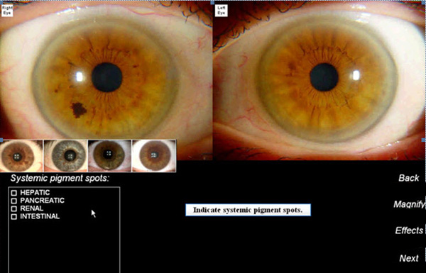
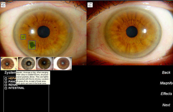
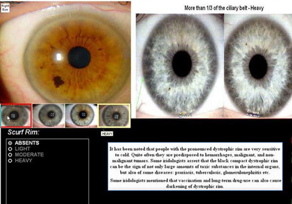
Morphologic Signs are important to determine chronic pathological conditions.
Scroll over iris samples in window to show more information on Systemic Spots.
Determine degree level of scurf rim.
6: Pupillary Belt & Collarette Indicators
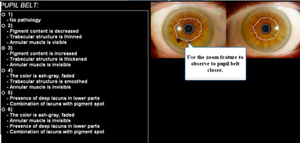
The Iridodiagnostic software includes analysis of changes of pupillary belt and collarette. These signs have been verified through several hospital clinical studies.
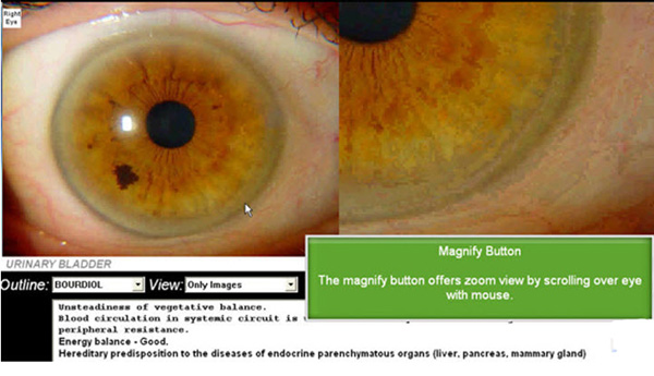
Scroll Mouse over specified eye to zoom in pupil sector.
Scroll mouse over text section and choose clinical analysis options.
6: Special Effects
The Iridodiagnostic software includes several types of special effects that can assist in analysis.
Sharpen
Gradient
Histogram
Importing/Capturing Iris Images
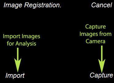
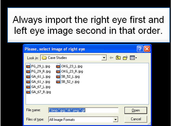
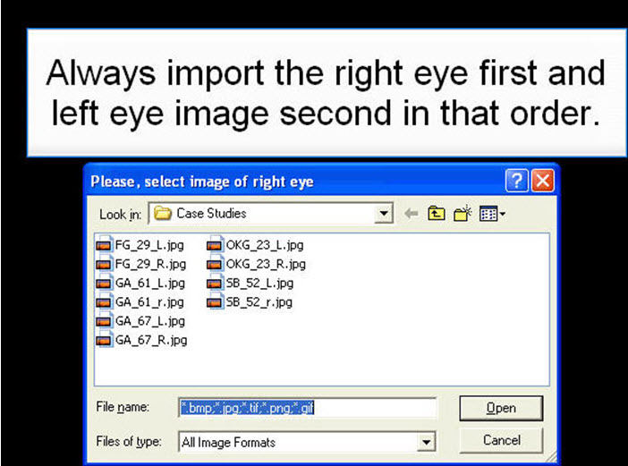
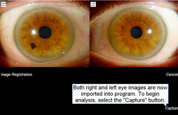
Images must be imported into program by clicking the import button. You can import both right and left eye images at the same time. Always import the RIGHT EYE image first.
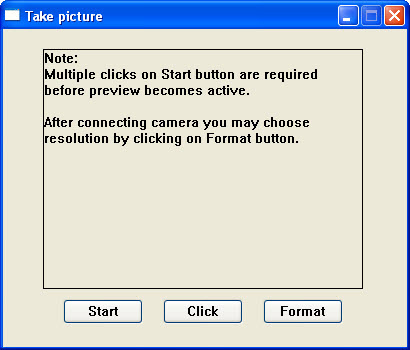
To capture an image for import, choose the capture button and click on the start button.
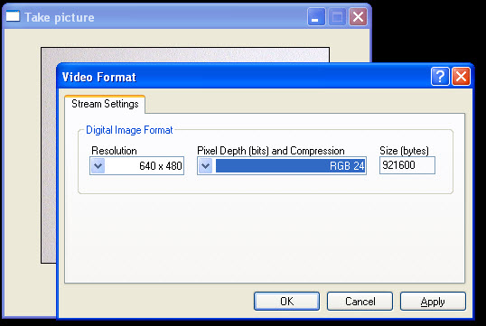
To capture images at different sizes, choose the FORMAT button. The best image size to use is 640X480
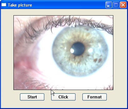
Use the click button to capture and store images.
Once you have captured and stored eye images for analysis, click the import button for importation of selected eyes for analysis.
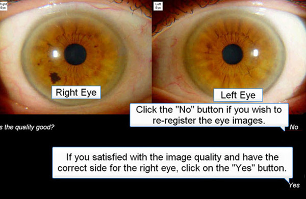
Once both right and left images are imported, choose the capture button:
If you are satisfied with the imported images, choose the ‘Yes’ button or ‘No’ button to re-import images.
Assessments
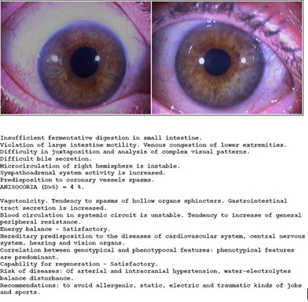
A final report assessment will automatically be generated after checking all the possible iris signs and parameters. The report can also be edited directly in program so that t
he user can modify data tailored toward their own clinical practice. If not familiar with certain terminology, they can refer to the included terminology guide. The assessment can be edited in the software before printing.

